



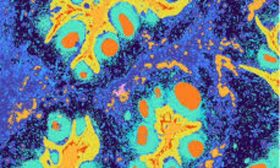
Multiple platforms for discovery are available for Spatial Transcriptomics.
Spatial Transcriptomics Services
10X
Takara
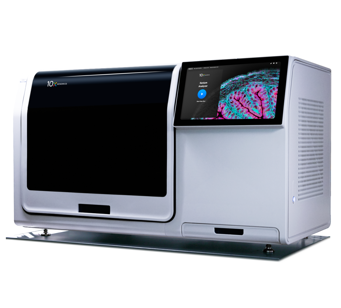
Xenium
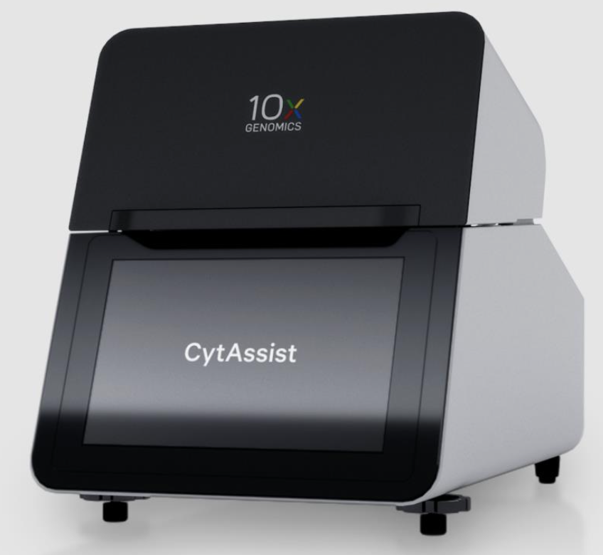
Visium
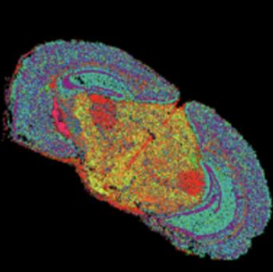
Seeker
Trekker
FAQ
Xenium
What is Xenium?
The Xenium platform offers an integrated solution for visualizing, quantifying, and analyzing gene expression and protein levels in fresh-frozen (FF) and formalin-fixed paraffin-embedded (FFPE) tissue sections at subcellular resolution. It features a range of validated and customizable gene panels, as well as fully custom panels, enabling sensitive profiling of specific genes of interest. The platform includes optimized Xenium Protein subpanels that facilitate multi-omics analysis of 27 markers related to the immune response and cancer microenvironments in human FFPE tissues, alongside panels covering fewer than 500 genes. A larger panel of 5,000 genes is also available. Data from the instrument can be transferred for further visualization and analysis using proprietary software or other third-party tools.
Do I need to stain my tissue?
It uses unstained tissue. Please consult with the core. Send an email to Emanuele Palescandolo, [email protected]
How big is the capture area?
Each run allows the capture of two sections of 12 mm x 24 mm. Multiple sections can be placed as long as they don’t overlap. Please consult with the core before starting. Send an email to Emanuele Palescandolo, [email protected]
What slides do I need to use?
The slides are proprietary and are provided by the core. Please send an email to Emanuele Palescandolo, [email protected]
What type of samples can I provide?
The instrument can analyze, fresh frozen and FFPE sections cut at 5 or 10 microns. Before starting any sample prep, please consult with the core. Send an email to Emanuele Palescandolo, [email protected]
How much does it cost?
The cost varies based on the panels nedeed. Please consult with the core. Send an email to Emanuele Palescandolo, [email protected]
Does it provide single-cell resolution?
The platform allows for the detection of RNA and protein in situ, therefore allowing for subcellular localization of the signal.
Visium
What is Visium?
10x Genomics Visium HD is a spatial transcriptomics platform designed for high-resolution mapping of whole-transcriptome gene expression across tissue sections at the level of individual cells. It employs a continuous grid of barcodes that provides millions of feature spots, enabling precise visualization of gene expression with spatial context in fresh-frozen, fixed-frozen, and formalin-fixed paraffin-embedded (FFPE) samples. The platform allows for comprehensive analysis of the entire transcriptome, ensuring complete coverage of tissue samples without gaps between capture areas. The technology is applicable to research in cancer, developmental biology, and complex tissue structures. The primary analysis is runwith the 10x Genomics proprietary software.
Do I need to stain my tissue?
The platform uses stained tissue to guide spatial mapping. Classic H&E or IF can be used, please consult with the core. Send an email to Emanuele Palescandolo, [email protected]
How big is the capture area?
Each run allows the capture of two sections (from 2 different slides) of 6.5 mm x 6.5 mm.
What slides do I need to use?
Sections must be provided on positively charged slides. Tissues that are prone to detachment may require specialized slides, please consult with the core. Send an email to Emanuele Palescandolo, [email protected]
What type of samples can I provide?
The instrument can analyze, fresh-frozen, fixed- frozen, and FFPE sections. Those need to be cut at 5 or 10 microns. Before starting any sample prep, please consult with the core. Send an email to Emanuele Palescandolo, [email protected]
How much does it cost?
The cost varies based on the tissue type. Please consult with the core. Send an email to Emanuele Palescandolo, [email protected]
Does it provide single-cell resolution?
The platform is not designed to identify cells and return their transcriptome, but the resolution is high enough to approach single-cell.
Seeker
What is Seeker?
Seeker Spatial Transcriptomics Kits facilitate complete transcriptome analysis and spatial mapping of fresh frozen tissues. This assay is compatible with any eukaryotic organism and does not necessitate the use of new specialized equipment or unique probe sets.
Do I need to Stain my tissue?
The method requires unstained, frozen tissue.
How big is the capture area?
The tiles come into 2 sizes, 3 mm x 3 mm and 10 mm x 10 mm.
What slides do I need to use?
None, the method requires sectioning tissues and placed directly on the proprietary tiles.
What type of sample can I provide?
Fresh Frozen tissue embedded in OCT to be cut at 10 micron. RNA QC should show an RNA integrity number (RIN) of at least 7. To increase the likelihood that the tissue structure to be captured is in a section, perform H&E or IF on some of the sections before choosing the unstained one for the assay. Please discuss with the core before submitting the material.
How much does it cost?
The cost is dependent on the number of reactions.
Does it provide single-cell resolution?
The beads microlayer provides a snapshot of the gene expression of your area of interest with a resolution of 10 μm, but it is not designed to be at single-cell resolution.
Trekker
What is Trekker?
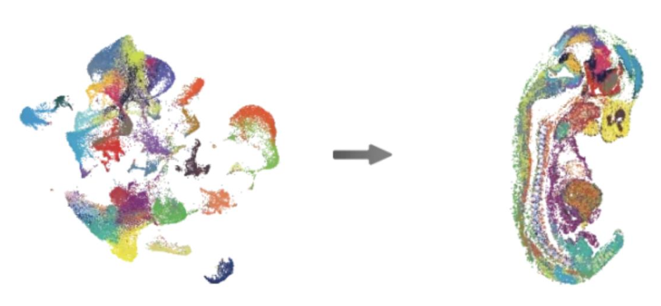
Trekker is a single-nuclei mapping kit, i.e., it integrates spatial profiling with single-nuclei data. It fits into existing single-cell sequencing workflows, allowing to move your UMAP clusters to a spatial map. It is a one-hour workflow before single-nuclei library preparation. It is compatible with virtually any organism.
Do I need to Stain my tissue?
The method requires unstained, frozen tissue.
How big is the capture area?
The tiles come in a 10 mm x 10 mm size.
What slides do I need to use?
None, the method requires sectioning tissues and place directly on the proprietary tiles.
What type of sample can I provide?
Fresh Frozen tissue embedded in OCT to be cut at 25-30 um micron. RNA QC should show an RNA integrity number (RIN) of at least 7. To increase the likelihood that the tissue structure to be captured is in a section, perform H&E or IF on some of the sections before choosing the unstained one for the assay. Please discuss with the core before submitting the material.
How much does it cost?
The cost is dependent on the number of reactions.
Does it provide single-cell resolution?
The method provides single-nuclei resolution.